Optical design#
Geometrical optics#
Geometrical optics or ray tracing allows approximation of light behavior as it passes through lenses, reflects off mirrors, or goes through interfaces between media with varying refractive index. It takes significant time commitment to absorb all topics of ray tracing and we are not aiming to do it here. Myriad of textbooks and other sources (see this StackExchange question) have been created that cover most of these details. Many papers that cover design of microscopy setups also provide explanation on principles involved, such as protocol for tiling light-sheet microscope or light-sheet light-field microscope design and large protocol collection from Nature, and sources from knowledge bases such as Thorlabs. However, here we would like to provide a brief description of potential topics and details that will aid in understanding microscopy systems and microscopy design.
Point-spread function (PSF)#
PSF represents a three-dimensional image of a point source and contains information about light wavelength, magnification, all types of aberrations, and spatial inhomogeneity of imaging system (e.g. PSF is different in center vs edge of image). PSF is tightly coupled with the definition of resolution: for two point objects to be “resolved” their PSFs need to be distinguishable. How we define that is up to the user, for example commonly used Rayleigh criterion sets distance between point sources at 0.61*wavelength/NA objective (see more here)
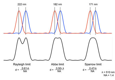
Image taken from UT Advanced Microscopy core
In super-resolution microscopy such as PALM/STORM/MINFLUX resolution is defined through statistical accumulation of estimated positions of point sources, not from single image. Use of deconvolution can mathematically increase resolution even though the optical system and corresponding PSF stay unchanged.
According to Niquist criterion to truthfully reconstruct PSF we need to sample it with at least twice the spatial frequency. If optical resolution (from PSF estimation) is expected to be 0.5µm, then we need to have data collection resolution 0.25µm (or finer) using scanning or camera-based detection.
Optical resolution necessary for particular experiment depends on the size of features we are interested in. As discussed in Designing microscopy experiments, lateral size and spacing between objects (molecules, cells, or regions of interest) is important to consider when designing experiment.
Aberrations and real optics#
We need to remember several common types of aberrations:
spherical: due to lens shape light at the edges is focused in different location that light along optical axis
chromatic: difference in light path of different wavelength due to dispersion
refraction within sample: due to non-homogeneity of refractive index of the sample (e.g. lipids vs proteins in the sample)
Some of these aberrations can be corrected by lens design, for example achromatic doublets can correct spherical and chromatic aberrations. Apart from price, these lenses can have higher reflection and absorption than comparable spherical singlets. Aspherical lenses are another alternative at even higher price and more limited
Designing and characterizing optical system#
If you can mathematically describe each element of the optical system you can perform design optimization and simulation to characterize expected performance. Foundational tool for that is proprietary Zemax however some free tools are also available for example Python-based Optiland
How to make things move#
kinematic mirror mount
Z stages
voice coils
linear actuators
piezo actuators
digital micromirror device (DMD)